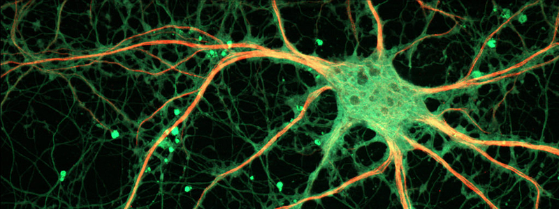
Multiple sclerosis is an inflammatory disease of the nervous central system leading to a progressive destruction of the myelin sheath surrounding the axons, essential for their protection and for the transmission of the nerve impulse. Bruno Stankoff and Catherine Lubetzki’s team developed a cutting-edge imaging technique which makes possible to visualize neuron demyelination and remyelination and to quantify their degeneration. These results, published in Annals of Neurology, open the way to better treatment for patients and to the evaluation of novel remyelination therapies.
Axones, neurons’ extension, are surrounded by a myelin sheath which plays a key role in the transmission of the nerve impulse and the protection of axons. In Multiple sclerosis, the immune system attacks the own elements of the patients and leads to the progressive destruction of the myelin sheath, also called demyelination, in the brain and the spine.
Multiple sclerosis is an autoimmune inflammatory disease. It is the most frequent demyelinating disease of the nervous central system and the first cause of non-traumatic severe disability in young adults. It leads to movement, sensitivity, balance, vision impairments…Multiple sclerosis affects about 100 000 individuals in France and more than 2, 8 million in the world. The disease affects women more than men, with a 3 for 1 ratio.
The quantification of demyelination and remyelination in MS patients is essential to better understand how does the disease progress and to evaluate possible remyelination therapies. Using positron emission tomography imaging or PET-SCAN (imaging technique), associated with specific molecules called tracers, could allow the observation of myelin dynamics in multiple sclerosis.
Scientists from Bruno Stankoff and Catherine Lubetzki’s team focused on two radiotracers: the [11C] PiB which can bind the myelin of the white matter, and the [11C] flumazenil, a molecule binding the neuron-specific GABA-A receptors.
They showed a progressive decrease in binding [11C] PiB between the undamaged white matter and white matter lesions caused by multiple sclerosis, reflecting a myelin decrease. Using [11C] PiB would allow to quantify the myelin dynamics in multiple sclerosis, i.e the demyelination and remyelination, and to categorize patients according to their ability to renew destroyed myelin sheathing, and direct the most appropriate course of treatment.
Researchers from Bruno Stankoff and Catherine Lubetzki’s team also looked at the degeneration of neurons in MS patients at different stages of the disease, using PET-SCAN with [11C] flumazenil.
Flumazenil is a molecule which can bind a receptor located on the synapse of neurons, the connection between two neurons and dendrites. They showed that flumazenil binding on its receptor decreases significantly in MS patients, with relapsing-remitting form in which relapses alternate with remitting phase with an improvement of the symptoms, as well as progressive forms in which there are no remitting phases.
These results open the way to a novel use of PET-SCAN with [11C] flumazenil to locate and quantify neurodegeneration in patients with multiple sclerosis. This quantification technique could also be used to evaluate neuro-protective drugs during clinical trials.
References :
Freeman L. et al. The neuronal component of gray matter damage in multiple sclerosis: A [11C] flumazenil positron emission tomography study. Annals of Neurology, 21 août 2015.
Bodini B. et al, Benzothiazole and stilbene derivatives as promising PET myelin radiotracers for multiple sclerosis, Annals of Neurology, 21 avril 2016.


