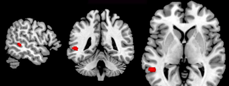
Researchers at the Institut du Cerveau – ICM recently showed for the first time that early lesions that are local-ized in a precise region of the brain occur 20 years before the appearance of frontotem-poral dementias. This major discovery could allow early diagnosis of the disorder and po-tentially lead to therapeutic trials and treating people at risk before they even develop clin-ical symptoms.
Frontotemporal dementias (FTD) are neurodegenerative disorders related to Alzheimer’s disease. They are nevertheless distinguished by the nature and location of lesions, which preferentially affect neurons of the frontal (anterior) and temporal (lateral) regions of the brain. These disorders manifest as behavior modifications such as apathy (loss of motiva-tion or taking initiative), disinhibition, difficulty with controlling emotions and eating be-havior, occurring around the age of 60.
Brain regions responsible for FTD, as with other neurodegenerative diseases, simmer quiet-ly for several years or decades before clinical symptoms appear. A major challenge for FTD research is to identify the region of the brain where the lesions start and to determine at what point they are formed.
In order to answer these questions, Drs. Isabelle Le Ber and Paola Caroppo, in the teams of Prof. Brice and Prof. Dubois are leading a study that aims to identify markers that will al-low diagnosis and early treatment for this disorder. This work is led in close collaboration with the neuroimaging teams of Drs. Marie-Odile Habert (LIB, UPMC) and Olivier Colliot (COGIMAGE, Institut du Cerveau – ICM) as well as with the Clinical Investigation Center of the Institut du Cerveau – ICM (Prof. Corvol).
Thanks to state of the art brain imaging techniques, the researchers identified a very small region of the brain, the middle temporal gyrus, that could be the origin of the first brain lesions in one genetic form of FTD. Structural anomalies (study of cortical thickness) and metabolic anomalies (via a positron emission topography study) are detectable in this re-gion around twenty years before the onset of the first symptoms of the disorder, showing that the lesion process is set in motion many years before the disorder manifests with clini-cal symptoms.
The identified region is involved in language comprehension (left hemisphere lesions) and also in the capacity to interact with others (social cognition) and to identify emotions (fa-cial emotion recognition), skills which are altered early in patients with FTD.
This major discovery allow localization of the beginning of the lesion process, in a form of the disorder, for a brain region before clinical appearance of the disorder. This region con-stitutes the first early lesion marker that could result in therapeutic trials and treating people at risk, in particular in genetic forms of the disorder, before patients develop the first clinical symptoms.


